Category: Cases/Blog

Home Category: Cases/Blog

The standard of care for the management of localized intermediate risk prostate cancer in Australia in most cases, involves treating the entire prostate. That is, either surgery to remove the prostate gland (robotic-assisted radical prostatectomy) or radiation therapy. For some men diagnosed with localised prostate cancer, there may now be another option. Focal therapy is an emerging treatment that targets only the area of the prostate containing significant cancer while preserving as much healthy prostate tissue as possible.

At Melbourne Urology Centre, our specialist Urologists can assess whether or not focal therapy is suitable for carefully selected men with localized prostate cancer. The goal is to effectively treat the cancer while reducing the risk of side effects such as urinary incontinence and sexual  dysfunction compared with surgery or radiation. Patients will undergo a comprehensive assessment, including prostate MRI, targeted biopsy and in most cases PSMA PET scan, followed by a discussion of all suitable treatment options.

melbourne urology team

Not every man diagnosed with prostate cancer require treatment and not all men be suitable candidates for focal therapy. The best results may be achieved in men with localised, clinically significant prostate cancer confined to a specific area of the prostate. Careful patient selection is essential to achieving good cancer control.

If you are seeking a second opinion or have recently been diagnosed with prostate cancer, the specialist Urologists at Melbourne Urology Centre can discuss whether focal therapy or other treatment including robotic assisted radical prostatectomy is appropriate for your individual situation. Patients from across Melbourne and Victoria are welcome to make an appointment for a consultation to explore personalised treatment options.

Who Is Suitable for Focal Therapy for Prostate Cancer?

Focal therapy is an emerging treatment option for carefully selected men with early stage localised prostate cancer. Rather than removing the prostate (robotic-assisted radical prostatectomy) or treating the entire prostate gland with radiation, focal therapy targets the area containing the clinically significant cancer while preserving as much normal prostate tissue as possible.

The best candidates for focal therapy are men with a single, well-defined area of intermediate-risk prostate cancer that can be accurately identified using high-quality prostate MRI and targeted prostate biopsy. In order to be considered for focal therapy, patients should have disease confined to the prostate, without evidence of spread elsewhere. Prostate cancer is often multifocal (that is, multiple areas of cancer within the prostate), which means not every patient will be suitable for focal therapy. For men with multi-focal, high volume or high-grade prostate cancer it is generally more appropriate to undergo treatment with established therapies such as robotic-assisted radical prostatectomy or radiation therapy. This is to ensure the best chance of cancer cure and to minimize the risk of leaving residual cancer or progression to metastatic disease.

A thorough assessment is essential before recommending focal therapy. This typically includes PSA testing, multiparametric MRI, targeted and systematic prostate biopsy, followed by staging PSMA PET scan and discussion about the most appropriate treatment options.

Patients need to understand that focal therapy remains an evolving treatment. Ongoing follow-up is required which involves regular PSA testing, repeat MRI surveillance as well as further prostate biopsies to ensure that the treatment has been effective. Close monitoring for recurrent or untreated cancer is an essential requirement for men undergoing focal therapy for prostate cancer.

In Australia, focal therapy is not currently funded by Medicare and is not covered by private health insurance, meaning there are out-of-pocket costs. If you are interested in discussing your suitability to undergo focal therapy for prostate cancer in Melbourne, a consultation with a specialist Urologist at Melbourne Urology Centre may help determine if you are an appropriate candidate for this treatment.

Kidney stones are known for causing severe discomfort, so understanding kidney stone pain location may help you to identify a problem early on so that you can seek medical attention.

Common Kidney Stone Pain Location: The Flank

The classic kidney stone pain location is often in the flank, which is the area on either side of your lower back, just below your rib cage.


Kidney stone pain in this location is caused by the ureter contracting in response to the presence of the stone(s). It is often described as “sharp” or “colicy”. It can come on quite suddenly, and usually goes in waves from 20-60 minutes of discomfort, before it stops and starts again.

Radiating Kidney Stone Pain Location

In addition to the flank, kidney stone pain location can radiate to other parts of the body. Many patients will report that their pain spreads to the lower abdomen and groin. In males, kidney stone pain can also be felt down to the testes.

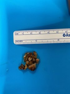
Fragmented kidney stone removed from the patient

Kidney Stone Pain Location and Urinary Symptoms

Kidney stone pain location is often also accompanied by urinary symptoms. Some patients may experience a frequent urge to urinate and/or pain with urination. There may also be changes in the colour of urine; it may appear pink, red, or brown, indicating the presence of blood.

Kidney Stone Pain Location: Kidneys vs Back Pain

Kidney stone pain location is sometimes confused with back pain. Distinguishing between the two is essential for your doctor to be able to make a proper diagnosis. While both conditions can cause pain and discomfort in the back area, kidney stone pain tends to be much more severe and persistent. Additionally, pain associated with kidney stone disease often shifts location and changes in intensity as the stone moves through the urinary tract, unlike typical back pain.

Kidney Stone Pain Location and Intensity

The severity of discomfort in the kidney stone pain location can vary depending on the size, position, and number of stones in the urinary tract. Even small stones can cause intense pain requiring urgent medical attention, if they obstruct the flow of urine.

Understanding Kidney Stone Pain Location and Managing It

Being aware of kidney stone pain location is the first step in managing kidney stone disease. Hydration is the most important factor in preventing kidney stones and their associated pain; having dilute enough urine can help to prevent any type of kidney stones and may help smaller stones to pass on their own.

In mild cases, over-the-counter pain medications may provide temporary relief, however persistent or severe pain should always be evaluated by your GP or urologist.

Kidney Stone Pain Treatment Options

The treatments that are offered by your urologist will be dependent on the size, number and location of kidney stones within your urinary tract. Your general health, lifestyle, and medical history will also be considered.

In milder cases, or where stones are very small and appear to be passing through the tract, certain medications may be prescribed to help manage pain and assist stone passage.

In cases of severe kidney stone pain, procedures like extracorporeal shock wave lithotripsy (ESWL) can be used to break down larger stones, while ureteroscopy with laser allows for the fragmentation and removal of stones using a small, specialised scope and basket.

Preventing Kidney Stone Pain and Recurrences

Once you have had a kidney stone, the risk of recurrence is up to 10% per year. Taking preventative measures is important in reducing recurrence. This includes staying hydrated and reducing sodium intake. In some instances, maintaining a diet low in oxalates may also help, however you should not go making drastic changes to your diet without your urologist’s advice. Having regular check-ups with your doctor may also help you monitor and manage your risk factors for kidney stone disease.

Kidney Stone Pain Location: When to Call a Doctor

Any time you are experiencing pain, you should seek medical attention. If you have any of the following symptoms, you should seek emergency medical care:

  • Pain that is severe, that comes on suddenly
  • +/- Difficulty or urgency to urinate
  • +/- Blood in your urine
  • +/- Fever/chills
  • +/- Vomiting

Kidney Stone Pain Location: The Bottom Line

Understanding kidney stone pain location is really important for early identification and effective management of painful stone disease. By recognising the specific areas where kidney stone pain often occurs, you can seek medical advice and treatment in a timely manner. If you experience any pain or urinary symptoms consistent with kidney stone disease, you should contact your GP or urologist for an accurate diagnosis and appropriate care plan.

Kidney stone symptoms in female patients are often very similar to those experienced by males, however they are sometimes overlooked or wrongly attributed to other causes in females.

Kidney stones are small, hard deposits of mineral and acid salts that form on the inner surface of the kidneys. In many instances, kidney stones do not cause any symptoms, however in cases where they are symptomatic, they can cause severe pain and discomfort.

This blog aims to explore the kidney stone symptoms in female patients and some causes, diagnosis, and treatment options.

What are Kidney Stones?

Kidney stones are small mineral deposits that form in the urinary tract. Stones may develop if the urine becomes too concentrated with certain substances. Types of kidney stones include calcium oxalate stones (most common), uric acid stones, struvite stones, and cystine stones. These present due to different causes and risk factors.

Kidney Stones Video:

Fragmented Kidney Stone Removed from Patient:

Fragmented kidney stone removed from the patient

Causes of Kidney Stones and Kidney Stone Symptoms in Female Patients

Before we take a look at kidney stone symptoms in females, we need to understand the causes of kidney stones.

The most common causes of kidney stones- and therefore kidney stone symptoms in female patients- include dehydration, some particular diets, obesity and some medical conditions. There are also some risk factors of women in general, such as pregnancy and the hormonal changes that come with it.

In general, a high-sodium diet combined with inadequate fluid intake and certain lifestyle factors also contribute to the overall risk of developing kidney stones.

General Symptoms of Kidney Stones vs Kidney Stone Symptoms in Females

As mentioned above, not all kidney stones cause symptoms; sometimes they are discovered incidentally. In cases where they do cause the patient symptoms, these can vary but often include:

  • Severe pain and discomfort in the side and back (flank), below the ribs, and usually radiating to the lower abdomen and groin.
  • Nausea and vomiting from the intense pain, or in cases where there is also infection present.
  • A need to urinate frequently and urgently, often in small amounts.
  • Blood in the urine, where blood appears pink, red, or brown.

Kidney stone symptoms in female patients may be slightly different. In addition to the general kidney stone symptoms, women may also experience:

  • Lower abdominal pain: whilst men often report back pain, women may experience more lower abdominal pain.
  • Symptoms during pregnancy: some women are at increased risk of developing kidney stones and unique symptoms due to changes in the urinary tract.
  • Menstrual cycle impacts: some women report that kidney stone symptoms can coincide with their menstrual cycle, making it harder to differentiate between menstrual pain and kidney stone pain.

Diagnosing Kidney Stones in Women

Kidney stone symptoms in female patients may vary slightly from those in males, however for the majority of cases, the diagnostic process is the same:

  • Physical Examination: the urologist will assess symptoms and medical history.
  • Imaging Tests: Ultrasound, CT scans, and X-rays may be used to locate and identify the size of the stones.
  • Urine and Blood Tests: These tests can detect high levels of minerals and other substances that cause stones.

Complications of Kidney Stones in Female Patients

Kidney stones that are symptomatic require treatment. If left untreated, kidney stones can lead to complications such as:

  • Urinary Tract Infections (UTIs): Kidney stones can block the flow of urine through the urinary tract, leading to painful inflammation and infection.
  • Kidney Damage: Persistent stones can cause damage to the kidneys due to pressure build up.

Treatment Options for Kidney Stone Symptoms in Female Patients

Treatment options for kidney stone symptoms in female patients are often the same as those offered to males, with this exception of pregnant women, who may require different treatments.

Treatment for each patient’s kidney stone symptoms will depend upon the size and type of stone and the severity of symptoms. Treatment is centered around:

  • Pain management medications
  • Medications to help stones pass through the urinary tract more easily
  • Surgical interventions such as ureteroscopy, lithotripsy (using shock waves to break up stones), and percutaneous nephrolithotomy (removing stones through a small incision) may be necessary for larger stones.

When to Seek Medical Attention

No matter whether it is suspected kidney stone symptoms in female or male patients, it is important to seek prompt medical attention for diagnosis. This is especially important if you are experiencing severe pain, blood in your urine, or symptoms of a urinary tract infection (fever, chills, nausea or vomiting). Early diagnosis can prevent complications and make treatment more effective.

Prostate cancer life expectancy…

If you or someone you know has recently been diagnosed with prostate cancer, then you might be wondering about – or indeed have some anxiety around – prostate cancer life expectancy.

We should begin this blog by noting that most men who are diagnosed with prostate cancer will not die from the disease.

Even so, prostate cancer remains an important health issue amongst Australian men. An insight into prostate cancer life expectancy is thus vital to make important decisions around treatment and care.

Today’s blog will take a look at the different factors that impact prostate cancer life expectancy, the survival rates by cancer stage, treatment methods and lifestyle considerations among others.


Prostate Cancer Video

What is prostate cancer?

So, what is prostate cancer? It’s just what it sounds like: a cancer that has developed in the prostate, a small gland found only in men which produces seminal fluid.

Prostate cancer has different forms:

  • Localised, or confined to the prostate
  • Locally advanced, in nearby tissues
  • Metastatic, which means it’s spread elsewhere in the body.

Stages of prostate cancer and how this affects prostate cancer life expectancy.

Prostate cancer life expectancy is affected by the stage of the cancer. Prostate cancer is categorised into stages based on how far it has spread:

  • Stage I: Cancer is confined to a small area of the prostate.
  • Stage II: Cancer is more advanced but still confined to the prostate.
  • Stage III: Cancer has spread outside the capsule of the prostate.
  • Stage IV: Cancer has spread to nearby structures and/or distant parts of the body.

Early diagnosis significantly affects prostate cancer life expectancy.

The stage of cancer at the time of diagnosis significantly affects prostate cancer life expectancy. Early detection and treatment can significantly improve a man’s chances of survival. Diagnostic methods for prostate cancer include the PSA (prostate-specific antigen) blood test, clinical examination and MRI-fusion transperineal biopsy.

Factors influencing prostate cancer life expectancy.

Several factors will influence prostate cancer life expectancy in a man. These factors include:

  • Gleason Score and Tumor Grade; Higher Gleason scores indicates more aggressive cancer and is the most important factor in determining prognosis
  • The man’s age at the time of diagnosis
  • The overall health of the man; Pre-existing health conditions can impact treatment options and outcomes
  • PSA level; In some cases, higher PSA levels can be associated with more advanced disease.

Early detection and treatment significantly improve survival rates and prostate cancer life expectancy.

Treatment options and their impact on prostate cancer life expectancy.

Your urologist will be able to offer different treatment options based on your individual circumstances, the stage and aggressiveness of the cancer:

  • Active Surveillance: This involves monitoring low-risk cancer closely with regular PSA testing, surveillance MRI and repeat prostate biopsy. Low risk cancer has an excellent life expectancy.
  • Surgery (Robotic-assisted Radical Prostatectomy): Removing the prostate surgically can be curative in early stage cancers, significantly improving prostate cancer life expectancy
  • Radiation Therapy: This option may be effective for localised and some locally advanced cancers, with good survival rates, similar to surgery
  • Hormone Therapy: This is sometimes offered in cases of advanced prostate cancer, to slow its progression and manage the symptoms, impacting life expectancy positively.
  • Chemotherapy and Targeted Therapies: These are often used for metastatic cancers to improve quality of life and extend survival.

Prostate cancer life expectancy and prognosis with lifestyle influences.

Lifestyle choices can affect the prognosis of cancer in general and overall life expectancy. These may include:

  • Diet and Nutrition: A healthy, balanced diet that is rich in fruits, vegetables, and low in red meat
  • Physical Activity: Regular exercise boosts overall health and may improve cancer prognosis
  • Smoking and Alcohol Use: Avoiding smoking and limiting alcohol can improve life expectancy and reduce overall cancer risk.
  • Mental Health and Support Networks: Psychological well-being and strong support networks are often just as important as the physical factors in improving quality of life in men with prostate cancer.

Prostate cancer life expectancy and living with prostate cancer.

For many men diagnosed with prostate cancer, living with their diagnosis involves managing treatment side effects and maintaining quality of life. This means making quality of life considerations when it comes to balancing treatment efficacy with any potential side effects. Your urologist, prostate cancer nurse, and support networks can help with strategies for managing side effects like fatigue, urinary issues, and sexual function. Regular monitoring by your urologist is important in order to quickly detect any recurrence and manage long-term health.

When to see your doctor.

The biggest factor in increasing prostate cancer life expectancy is early detection and treatment. All men over the age of 50 who are interested in assessing their risk of prostate cancer should visit their doctor for a PSA blood test. For men with a family history of prostate cancer, this may be performed from age 45. Similarly, anyone experiencing symptoms such as difficulty urinating, blood in the urine, or pelvic discomfort should consult with their family doctor.

Prostate cancer life expectancy: the bottom line.

Whilst a diagnosis of prostate cancer can be daunting, it is important for patients and their families to have an idea of what the prostate cancer life expectancy looks like. With the guidance of their urologist, patients and their families are able to gain an understanding of the factors that affect prognosis, and consider various forms of treatment that are appropriate for their circumstances.

Renal colic is a sudden and severe form of pain in the flank, caused by kidney obstruction. Often, renal colic is caused by an obstruction of the urinary tract by a kidney stone.

Renal colic treatment is tailored to the severity of pain, underlying cause, patient’s overall assessment.  The initial management is to alleviate the pain efficiently and then address the underlying cause.

Renal colic treatment: What is renal colic?

It is estimated that renal colic is experienced by 5-15% of the population most often due to a kidney stone. Renal colic, or acute kidney pain, is characterised by excruciating flank pain caused by a kidney stone blocking the urinary tract. Kidney stones form when minerals/salts in the urine clump together, creating crystals, which can then become lodged in the urinary tract. If a stone blocks the urinary tract and restricts the flow of urine, this can cause a build up of pressure and inflammation.

Kidney stones can form in the urinary tract for various reasons, including:

  • Dehydration
  • Excess calcium in the urine
  • Excess protein in the diet
  • Certain medications
  • Hyperparathyroidism
  • Some gastrointestinal conditions, such as Crohn’s Disease or ulcerative colitis.

Renal colic treatment is based on prompt and effective action to alleviate pain and prevent complications. The renal colic treatment approach that our urologists use encompasses various modalities that are tailored to the patient’s condition and stone characteristics.

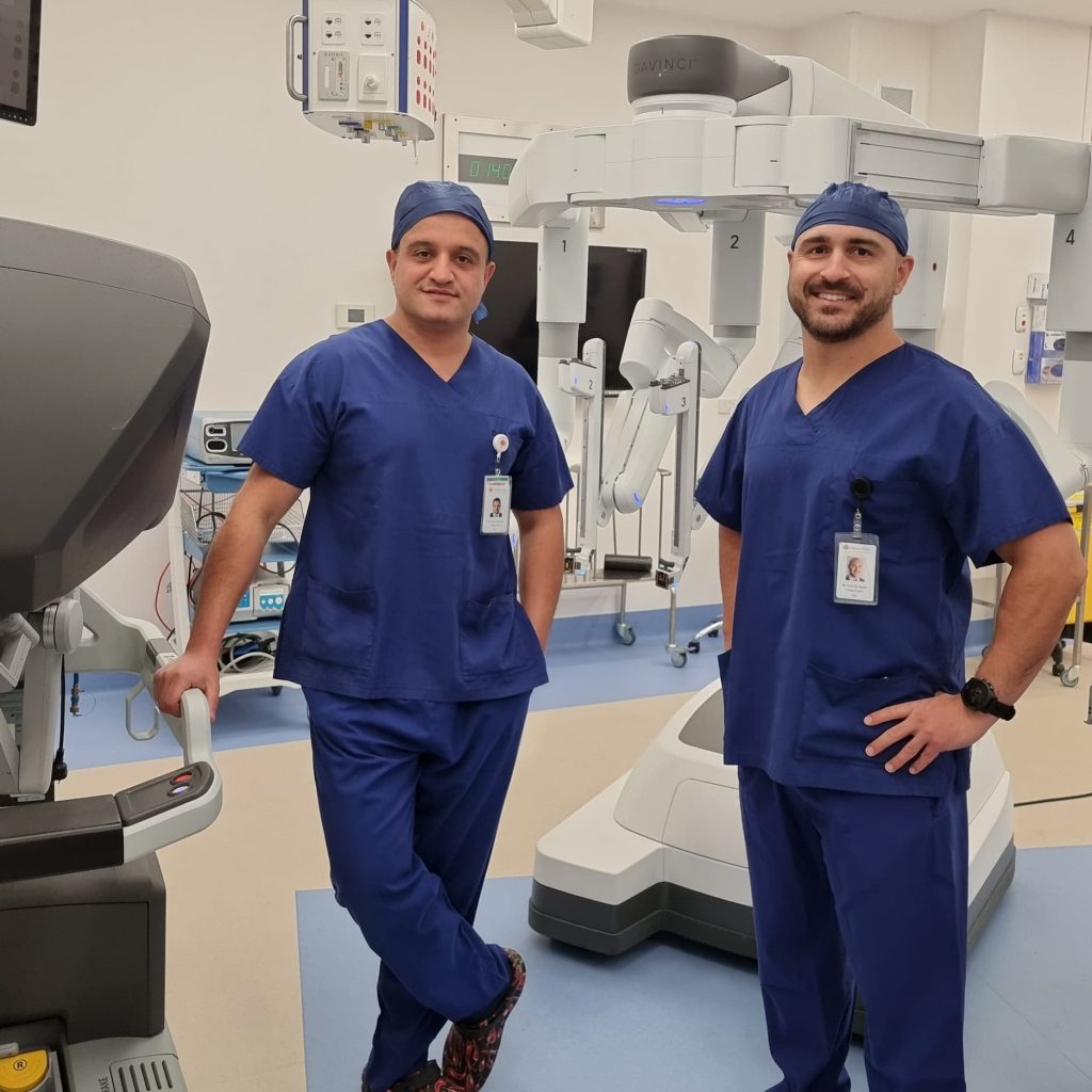
best urologists melbourne shekib and tony

Renal colic treatment: Immediate intervention

Immediate renal colic treatment usually focuses on pain relief and antiemetics (anti-nausea drugs) to address the acute pain and associated symptoms such as nausea. Nonsteroidal anti-inflammatory drugs (NSAIDs) and opiates are often at the cornerstone of analgesic (pain relief) therapy. NSAIDs are useful as not only do they alleviate pain, but they also reduce pressure in the kidneys, offering dual benefits.

Rehydration is also important during the immediate stages of renal colic treatment. Many patients become dehydrated due to vomiting or simply reduced fluid intake whilst they are feeling unwell.

Renal colic treatment: Dealing with the stone

The next phase of renal colic treatment is to begin the process of moving or removing the obstructing stone. Alpha-blocker medications like tamsulosin or nifedipine, can help with stone passage by reducing the pressure inside the ureters. While their efficacy varies between patients, they may be of benefit to patients with smaller, distal stones.

For impacted stones that require definitive management, more surgical procedures such as shock wave lithotripsy or ureteroscopy may be suggested by the urologist.

Your urologist may also recommend that a ureteric stent be placed. This will help to allow the urine to drain more freely and therefore assist with relieving the pressure in the urinary tract. Ureteric stents also prepare the ureter for a safe passage of a ureteroscope and lasering of the ureteric/kidney stone.

Renal colic treatment: Timely intervention is important

If you experience sudden, severe kidney pain, it is important that you contact your doctor right away, even if there is no blood in your urine.

Timely intervention is important in renal colic treatment, especially for obstructing stones. The sooner you seek medical help, the better chance you will have of reducing complications.

As part of your longer-term renal colic treatment, your urologist may recommend some dietary changes and increased fluid intake, depending on the types of kidney stones present.

Renal colic treatment: In conclusion

Successful renal colic treatment is based on a comprehensive approach that addresses pain relief, hydration, stone passage (or definitive interventions), dietary or fluid modifications, and possibly other preventative measures.

At Melbourne Urology Centre, our urologists are experts in renal colic treatment. They work closely with their patients to tailor an effective approach that alleviates symptoms, mitigates complications, and empowers patients in preventing stone recurrence.

We understand that a diagnosis of prostate cancer can be a daunting prospect, but we believe that understanding the process of how prostate cancer is diagnosed is important for our patients, as it can help ease anxiety and empower patients to be actively involved in making informed decisions about their health.

Today’s blog looks at how prostate cancer is diagnosed, exploring the methods and tests that we use to make a definitive diagnosis.

How prostate cancer is diagnosed: Causes and risk factors

When we discuss how prostate cancer is diagnosed, we firstly must consider the risk factors. Whilst there are no definitive causes of prostate cancer in men, there are some known risk factors:

  • Age, increasing risk especially after 50 years old
  • A family history of prostate, breast or ovarian cancer (especially with BRCA1 and BRCA2 gene mutations)
  • A first degree relative (father or brother) diagnosed with prostate cancer before the age of 60
  • There may also be an association with high testosterone levels.

Men who have one or more of these risk factors should consider speaking to their doctor about how prostate cancer is diagnosed, and discuss prostate cancer screening.

How prostate cancer is diagnosed: Could it be cancer?

Prostate cancer affects around one in seven Australian men. Whilst most men won’t die from the disease, it remains a leading cause of death in Australian men.

Having an understanding of how prostate cancer is diagnosed can help; early detection and treatment can improve survival from prostate cancer.

Early prostate cancer does not usually cause any symptoms. Some of the symptoms of advanced prostate cancer may include:

  • Frequent and urgent need to urinate
  • Difficulty or pain during urination
  • Blood in the urine or semen
  • Weak urine stream
  • Pain in the back or pelvis, or other areas of the body (if the disease has spread to axial bones or other parts of the body).

It is important to note that these symptoms can also be caused by a number of other conditions, so experiencing one or more of these does not necessarily mean that prostate cancer is present.

The only way to definitely diagnose prostate cancer is by conducting a series of tests specifically for it. If your urologist suspects that you may have early or advanced prostate cancer, it may be recommended that you have some of the tests that we will describe below.

How prostate cancer is diagnosed: PSA Blood testing

The PSA (prostate specific antigen) test measures the level of PSA, which is a protein that is made by both normal and cancerous cells of the prostate. Whilst the PSA blood test does not definitely diagnose prostate cancer, it is a useful tool for your doctor, as it can be used to assess changes in your PSA levels over time, helping to determine your risk of prostate cancer.

How prostate cancer is diagnosed: MRI fusion transperineal biopsy

When we’re looking at how prostate cancer is diagnosed, prostate biopsy is the only definitive way to obtain a definitive diagnosis.

MRI fusion transperineal prostate biopsy is the most accurate and comprehensive way to sample the prostate for changes that may be related to prostate cancer or other conditions.

An MRI fusion prostate biopsy is used to remove small samples of tissue from the prostate gland. For this procedure to take place, the patient will have had an MRI scan prior. The MRI image is fused with a live ultrasound image, generated through an ultrasound probe that is inserted into the patient’s rectum. A special biopsy grid is positioned against the skin behind the scrotum. This allows the surgeon to collect biopsies from specific areas of interest in the prostate.

The samples are sent off and examined by a laboratory. If prostate cancer is detected, the biopsy samples will also be used to determine the aggressiveness of the disease (Gleason score). This information can then be used to guide treatment recommendations.

How prostate cancer is diagnosed: In Conclusion

When considering how prostate cancer is diagnosed, it is important to remember that the process involves a comprehensive approach by the urologist. This approach combines the patient’s medical and family history, a physical examination, imaging tests, biopsies, and laboratory analyses.

The urologists at Melbourne Urology Centre will be able to guide you through the diagnostic process, working closely with you to detect any abnormalities and determine an appropriate treatment and management plan.

By seeking prompt medical attention for those who are at higher risk of developing prostate cancer, or in symptomatic cases, patients can improve their chances of early detection and timely intervention.

What is prostate cancer screening?

Prostate cancer screening involves conducting a PSA blood test. This test measures the amount of prostate-specific antigen (PSA) in the blood. Every man will have a level of PSA detectable in their blood, as the PSA protein is produced by normal, as well as by malignant cells in the prostate gland.

The PSA test is not a specific cancer test, but rather a prostate test, forming one component of the prostate cancer screening process. In men with prostate cancer, the PSA reading will usually be higher than in those without. The aim of the PSA test is to detect prostate cancer at an early stage, so that the chances of curing the disease are improved.

What is prostate cancer and why is screening important?

The prostate is a walnut-sized gland that sits just below the bladder in men. Its function is to produce some of the components of semen. When the cells of the prostate gland grow in an abnormal and unregulated manner, prostate cancer occurs. There are different types of prostate cancer. Some types of cancers grow slowly, rarely causing the patient any harm, whilst others can spread to other parts of the body (metastasize), causing serious harm to the patient, and sometimes these can even result in death.

Most men who are diagnosed with prostate cancer won’t die from the disease. Still, prostate cancer is the cause of the second highest number of cancer-related deaths in Australian men, and as such it is a major health concern.

Prostate cancer screening is important, because prostate cancer rarely causes a man to experience any symptoms until it reaches an advanced (and often incurable) stage.

Is prostate cancer screening necessary for all men?

Prostate cancer screening has been scientifically proven to save lives. A large-scale European study with over 160,000 participants showed that PSA screening led to a 20% reduction in death from prostate cancer in men who were screened.

Despite this evidence, there has unfortunately been some controversy around the PSA screening test. This is largely due to the fact that PSA screening has the potential to detect low-grade tumours which would otherwise be undetected, never cause any symptoms, and would not generally require treatment. This may cause anxiety in some patients.

This is where the role of the urologist is particularly important in the prostate cancer screening process: the urologist will educate and manage the patient appropriately to avoid over-treatment in these cases.

PSA screening/prostate cancer screening remains an effective way to diagnose high-grade prostate cancers at an early stage, allowing them to be treated in a timely manner, giving the patient the best chance of cure.

Who should undergo prostate cancer screening?

Making a decision as to whether or not to undergo prostate cancer screening is a personal choice. The major benefit of undergoing prostate cancer screening is early detection, with improved chances of timely treatment and cure.

Men who are interested in determining their individual risk of having or of developing prostate cancer should speak to their family doctor about having a prostate cancer screening blood test (PSA blood test).

The general recommendation for prostate cancer screening for men is:

  • At 50 years of age, if there is no family history of prostate cancer
  • At 45 years of age if there is a first degree relative (father, uncle or brother) with prostate cancer.

How is a prostate cancer screening conducted?

Prostate cancer screening usually begins with the collection of a blood sample from the patient, to conduct a PSA blood test. The sample is sent to the laboratory for analysis. Results are usually reported as nanograms of PSA per millilitre (ng/mL) of blood.

It is important for patients to understand that there is no ‘normal’ level of PSA in the blood. As mentioned earlier, PSA will be detectable in the blood of all men who still have their prostate. There are a number of conditions which are not cancer such as an enlarged prostate (BPH), inflammation (prostatitis/ infection) that may also cause the PSA level to be elevated.

This is the main reason why the PSA blood test only forms part of the prostate cancer screening process. It also highlights the importance of monitoring PSA levels over time, rather than considering only a standalone reading.

What happens if the PSA is elevated on the prostate cancer screening test?

If the reading on the PSA screening test is elevated, generally your doctor will ask you to repeat the test in 4-6 weeks. This will help your doctor to determine whether or not it was a true reading. From there, you will be referred to a urologist, who will perform a comprehensive assessment to determine your individual risk. Your urologist will perform an examination and may recommend further investigation with a multiparametric MRI and transperineal prostate fusion biopsy. This procedure can definitively diagnose or exclude prostate cancer.

If a diagnosis of prostate cancer is made, then your urologist will ensure that you are fully informed of all of your options for management of your condition, including active surveillance in cases of low-grade, low-risk cancers, to avoid over-treatment.

Prostate enlargement is also sometimes referred to as BPH, or benign prostatic hyperplasia. It is a common condition that affects aging men. With increasing age, the prostate gland can undergo non-cancerous growth. This can cause troublesome urinary symptoms for some men.

If you have been diagnosed with BPH, you might be wondering what prostate enlargement treatment options are available. In this blog, we will cover some of the available options, however the best source of information is always your urologist, who will be able to advise you which treatments are the most appropriate for your condition.

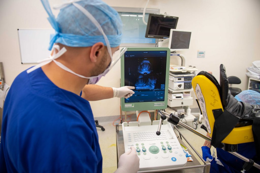
Dr Tony de Sousa examining an image from the MRI fusion prostate biopsy

Prostate enlargement treatment options– What is the prostate gland?

Before we can look at prostate enlargement treatment options, it is important to understand what the prostate gland is and where it is located. The prostate is located just below the bladder, in front of the rectum. It is about the size of a walnut and forms part of the urethra (the tube through which urine flows out of the body). The prostate produces part of the fluid that makes up semen.

In some men, when the prostate becomes enlarged, the tissue becomes obstructive and causes bothersome urinary symptoms. These symptoms can include difficulty starting/stopping urination, slow or dribbly flow, increased frequency and urgency to urinate, and frequent waking in the night to urinate (nocturia).

Thankfully, there are a number of effective prostate enlargement treatment options available.

Prostate enlargement treatment options– Medical treatments

Medical therapy is often one of the first prostate enlargement treatment options that men opt for. Medical therapy means the use of pharmaceutical medications including alpha-blockers and 5-alpha reductase inhibitors. These are the two main classes of drugs used in the treatment of prostate enlargement.

Alpha-blockers: This class of drugs works by relaxing the muscles in the prostate and bladder neck. This allows urine to flow more freely, thereby reducing the symptoms caused by the enlarged prostate gland. Some commonly prescribed types of alpha-blockers include tamsulosin, silodosin and prazosin.

5-alpha reductase inhibitors: These medications work by blocking the conversion of the hormone testosterone to dihydrotestosterone (DHT). DHT is known to contribute to the growth of the prostate. Finasteride and dutasteride are two examples of 5-alpha reductase inhibitors that may be prescribed.

For some men, combination therapy using medications that contain both alpha-blockers and 5-alpha reductase inhibitors is the most effective prostate enlargement treatment, in terms of medical options.

Prostate enlargement treatment options– Minimally invasive procedures

Minimally invasive procedures are another option for prostate enlargement treatment. Men may choose this option when medication does not provide them with adequate relief from their symptoms, or perhaps they do not wish to take medication in the long term (possibly due to side effects, cost, or other reasons). Additionally, minimally invasive procedures may be appropriate to consider in circumstances where the patient’s symptoms worsen.

Some of the common minimally invasive prostate enlargement treatment options include:

TURP (transurethral resection of the prostate): The patient is placed under anaesthesia, and a special instrument known as a resectoscope is inserted through the urethra. The obstructing prostate tissue is then removed. The TURP procedure can provide significant relief from symptoms for men with BPH.

Rezum therapy: this prostate enlargement treatment uses thermal energy (steam) from water vapour to ablate excessive prostate tissue, which causes it to shrink. The procedure is performed under anaesthetic. Most patients find significant improvement in their urinary symptoms following Rezum therapy. In the majority of patients, sexual function is preserved.

Other minimally invasive prostate enlargement treatments: There are some other options for minimally invasive treatments for BPH as well. These include therapies such as laser (HoLEP or greenlight) and Urolift. You can speak to your urologist about these treatment options, or find more information under the ‘Procedures’ tab on our website.

Prostate enlargement treatment options– robotic enucleation of the prostate

Robotic enucleation of the prostate is a treatment that may be offered in instances where the prostate is too large for TURP, laser or minimally invasive procedures.

Robotic-assisted enucleation of the prostate replicates the traditional open surgery, but instead uses the Da Vinci Robotic Surgical System to assist the surgeon, facilitating the use of very small robotic instruments to remove the obstructive tissue as a keyhole procedure.

Prostate enlargement treatment options to discuss with your urologist

Once a diagnosis of BPH has been made, you’ll want to know what your prostate enlargement treatment options are. BPH can have a profound impact on a man’s quality of life, so speaking to your doctor about early diagnosis and prompt treatment is essential in order to effectively manage the condition.

Fortunately, there are a number of different types of prostate enlargement treatment options available, ranging from medical therapies, through to minimally invasive procedures and robotic surgery.

If you are experiencing urinary symptoms, speak to your GP or consult a urologist for an accurate diagnosis. If you are diagnosed with BPH, your urologist will be able to recommend the treatments that will be most effective for your individual condition.

At Melbourne Urology Centre, our urological surgeons are highly experienced in prostate enlargement treatment. With our specialised care, men suffering from urinary symptoms can find prompt and effective relief, and lead a more comfortable life.

If you have been diagnosed with either a cancerous or benign (non-cancerous) tumour on the kidney, your urologist may have recommended that you have a partial nephrectomy. In this article, we’ll answer the question of ‘what is a partial nephrectomy?’ and explain the procedure.

What is a partial nephrectomy?

So, what is a partial nephrectomy? It is a surgical procedure during which the urological surgeon removes a cancerous or benign tumour from the kidney. This leaves the healthy kidney tissue in place, and so sometimes this procedure is also referred to as a kidney-sparing procedure.

Sometimes a partial nephrectomy may be performed to treat other kidney conditions as well, such as removing the damaged part of a kidney following trauma or injury.

Partial nephrectomy is considered a minimally-invasive procedure. At Melbourne urology Centre, our urological surgeons usually perform this procedure with the assistance of the Da Vinci Surgical robot, through keyhole incisions.

Partial nephrectomy allows for the preservation of the healthy part of the kidney tissue, so patients have a lower risk of kidney failure in the longer term.

What is a partial nephrectomy and when is it performed?

A partial nephrectomy is most commonly performed to remove a tumour from the kidney. This type of procedure is generally most appropriate for tumours that are 4cm or less in diameter, however, on occasion larger tumours of 5-7cm can be removed from the kidney via this method, depending upon their position on the kidney.

In patients with larger kidney tumours, undergoing a partial nephrectomy may not be a feasible or safe option. In these cases a complete (radical) nephrectomy is usually indicated.

Partial nephrectomy might also be performed in cases where a patient has scarred, damaged, or otherwise non-functioning kidney tissue, perhaps due to illness or traumatic injury.

What is a partial nephrectomy and how is it performed?

At Melbourne Urology Centre, our urological surgeons use the da Vinci robotic surgical system to perform most partial nephrectomy procedures.

Our urologists, Dr Tony de Sousa and Dr Shekib Shahbaz, are fully trained and highly experienced in performing robotic-assisted surgeries. The robotic device is under the control of the surgeon at all times. The use of the da Vinci robot offers numerous benefits to both the patient and the surgeon.

Partial nephrectomy is performed under general anaesthetic. The operating time varies from case to case, but generally the procedure takes 3-4 hours to complete.

What is a robotic partial nephrectomy and what happens during the procedure?

During a partial nephrectomy, keyhole incisions are made in the patient’s abdomen. The robotic instruments and specialised camera are inserted through these incisions.

Under the surgeon’s control, the robotic instruments are used to carefully and precisely remove the kidney tumour or damaged kidney tissue, preserving the normal kidney tissue. When the tumour has been removed, the kidney is meticulously repaired and the skin incisions are closed using plastic surgery techniques, for minimal scarring.

What is a robotic partial nephrectomy and what are the advantages of this type of procedure?

Performing the partial nephrectomy robotically offers numerous benefits both to the patient and to the surgeon.

The da Vinci robotic surgical system offers the surgeon:

  • High definition 3D vision inside the patient’s body
  • A greater range of motion and dexterity than the human hand, allowing for very precise movements
  • Overall greatly enhanced vision and precise control of the surgical instruments.

The da Vinci robotic surgical system offers the patient:

  • Less trauma: incisions are small and inconspicuous
  • Shorter blood cut-off (ischaemia) time: the kidney’s blood supply is restricted for less time with robotic partial nephrectomy, compared with conventional laparoscopy, which reduces the risk of long-term kidney damage
  • Less blood loss: there is less blood loss associated with robotic partial nephrectomy compared with traditional surgery, due to smaller incisions and greater precision
  • Faster recovery time: patients who have robotic-assisted surgery generally have a shorter hospital stay and a faster recovery compared with those who undergo conventional surgical methods.

What is a robotic partial nephrectomy and what can patients expect following the procedure?

Most patients will need to stay in hospital for 2-3 nights following their procedure. There will be some mild pain or discomfort, but this is generally well managed with medications. Patients are encouraged to sit out of bed and move around, as well as to perform deep breathing exercises in order to help reduce the risk of pneumonia or blood clots in the legs or lungs (DVT).

Most patients find that they are comfortable enough to return to their normal activities within 2-3 following having a partial nephrectomy. Strenuous activity and heavy lifting should be avoided for 6 weeks following the procedure to minimize the chances of bleeding.

Your surgeon will provide you with comprehensive post-operative instructions following your surgery.

A diagnosis of kidney cancer can be a daunting prospect, but we believe that understanding the process of how kidney cancer is diagnosed can help ease anxiety and empower our patients to be involved in making informed decisions about their health.

In this blog, we will take a look at how kidney cancer is diagnosed, exploring the various methods and tests that we, as urologists use to make a diagnosis.


urology specialist - best urologists


How kidney cancer is diagnosed: Could it be cancer?

It is worthwhile mentioning that most kidney cancers are discovered incidentally (by chance) when a person has an ultrasound or other imaging scan for an unrelated reason.

In cases where symptoms occur, they can include:

  • Blood in the urine or change in the colour of the urine (appears dark, rusty or red)
  • A lump in the abdomen
  • Pain the lower back that is not related to injury
  • Constant tiredness
  • Unexplained weight loss
  • Fever that is not caused by cold or flu.

These symptoms can also be caused by a number of other illnesses, so experiencing one or more of these does not necessarily mean that there is kidney cancer; the only way to definitely diagnose kidney cancer is by testing specifically for it.

If your urologist suspects that you may have an early or advanced kidney cancer, it may be recommended that you have some of the following tests.

How kidney cancer is diagnosed: Blood and urine tests

Whilst these tests don’t diagnose kidney cancer, they do check your general health and look for signs of problems with the kidneys. Blood and urine tests might also help to rule out certain conditions. Tests can include full blood cell counts (red cells, white cells and platelets) and tests to check how well the kidneys are functioning.

Urinalysis can detect the presence of blood or abnormal proteins in the urine, which may be signs of kidney cancer. Additionally, urine cytology may be performed to examine for cancer cells in the urine.

How kidney cancer is diagnosed: Imaging tests

To get a better look at the kidneys and surrounding areas, several imaging tests may be recommended by your urologist. These may include:

  • Ultrasound: This test is non-invasive test. A handheld probe is used to direct high-frequency sound waves to create images of the kidneys. It can help detect tumours in and around the kidneys and provide an initial assessment of their size and location. Ultrasound is painless and usually takes around 15 minutes to complete.
  • CT Scan (Computed Tomography): If a kidney cancer is suspected on an ultrasound, a CT scan may be recommended. A CT scan provides more detailed cross-sectional images of the kidneys and surrounding structures. It is especially useful in determining the size and extent of kidney tumours. It is painless and takes around 30-40 minutes. A CT scan can also be helpful in determining whether cancer has spread to nearby lymph nodes.
  • MRI Scan (Magnetic Resonance Imaging): This type of scan uses magnetic fields and radio waves to produce highly detailed cross-sectional images of the kidneys. It can help assess the tumour’s location and whether it has spread to nearby lymph nodes or organs. It can take between 30-90 minutes.
  • PET Scan (Positron Emission Tomography): PET scans use an injection of a small amount of radioactive solution that makes cancer cells show up brighter on the scan. This type of scan is useful for some cancers only.

How kidney cancer is diagnosed: Biopsy

Kidney biopsy is a definitive way to diagnose kidney cancer.

If your urologist suspects that there is a kidney cancer present, in some cases, they may recommend a kidney biopsy. During a biopsy, a small sample of tissue is removed from the kidney using a thin biopsy needle. This tissue is then examined under a microscope to confirm the presence of cancer and determine its type and aggressiveness. Kidney biopsies are typically performed under anaesthetic and guided by imaging techniques (such as ultrasound) to ensure precision and safety. This is not routinely required in all cases of kidney cancer.

How kidney cancer is diagnosed: Staging and Grading

Once a diagnosis of kidney cancer has been made, your urologist may order further testing in order to determine the stage and grade of the cancer. Staging assesses how far the cancer has spread, while grading evaluates how aggressive the cancer cells appear under a microscope. This information is important for preparing an appropriate treatment plan.

How kidney cancer is diagnosed: In Conclusion

When considering how kidney cancer is diagnosed, it is important to remember that the process involves a comprehensive approach that combines medical history, physical examination, imaging tests, biopsies, and laboratory analyses. Your urologist will be able to guide you through the diagnostic process and recommend which tests are appropriate for your circumstances.

By seeking prompt medical attention in symptomatic cases, patients can improve their chances of early detection and timely intervention.

Request an Appointment

We are here to assist you before, during and after your visit to the Melbourne Urology Centre. To request an appointment, simply complete the form below and we will contact you within 48 hours to confirm your appointment with us. For urgent enquiries or consulations please contact us on 1300 702 811