Category: Case Study

Home Category: Case Study

Mr. Jones is a 58 year old gentleman who was referred to Melbourne Urology Centre with bothersome urinary symptoms. Mr. Jones had been experiencing these symptoms for quite some time, however they were becoming increasingly frustrating to deal with and impacting his quality of life. With some encouragement from his wife, he made an appointment with his GP, who ran some preliminary tests before referring him to consult with a urologist.

Mr. Jones was diagnosed with benign prostatic hyperplasia (BPH), and following consultation with the urological surgeon, opted for Rezum treatment.

Symptoms and initial GP assessment

The symptoms that Mr. Jones presented with were very common to male patients with benign prostatic hyperplasia (BPH), also sometimes referred to as prostate enlargement. These urinary symptoms included:

  • Straining and difficulty to begin urination
  • Weak urine stream
  • Stop/start flow of urine
  • Urgency to urinate
  • Frequent urge to urinate overnight (nocturia), causing disturbance to his and his wife’s sleep.

Mr. Jones’ GP performed initial tests which included a urine test, PSA blood test and ultrasound. The urine sample was normal, with no blood or signs of infection. PSA (prostate-specific antigen) test was 1.2- normal for a man of his age. The ultrasound showed an enlarged prostate, measuring 60cc (normal size ~30cc) with incomplete bladder emptying.

The GP suggested a trial of medication. Tamulosin was prescribed, however this caused undesirable sexual dysfunction for this patient. Mr. Jones was then referred to Melbourne Urology Centre.

Consultation with a urologist

During the consultation with the urologist, a thorough assessment of Mr. Jones’ condition was performed. This included examination of the patient’s medical and family history. Mr. Jones did not have a history of prostate cancer in his family, however his father did have an enlarged prostate. The urologist assessed the prostate, and no sign of cancer was detected.

A flow test was performed at the urologist’s rooms, which showed slow flow and incomplete emptying of the bladder.

Rezum treatment for BPH

All of the treatment options were discussed with Mr. Jones. The patient noted that he preferred not to take medication.

Mr. Jones opted for Rezum therapy for prostate enlargement, due to the fast recovery time, less bleeding and less risk of sexual dysfunction than traditional surgery. He had an uncomplicated procedure, stayed overnight in hospital and was discharged the following morning.

Outcomes of Rezum treatment for prostate enlargement

Mr. Jones noted an improvement in his urinary symptoms after a few weeks, with better urine flow and bladder emptying. As expected, the bladder symptoms of frequency and urgency to urinate improved after a few months with bladder retraining, as guided by the urologist.


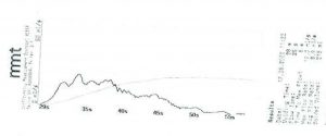
Flow rate results following Rezum therapy indicating a maximum flow of 19 mL/sec.

If you would like to discuss Rezum treatment for prostate enlargement or other treatments for BPH, please speak to your GP and obtain a referral for one of our urologists at Melbourne Urology Centre.

This case study is unique to the individual patient as described and the name has been changed to ensure patient confidentiality. Treatment details should not be taken as general medical advice. If you are suffering with BPH or urinary symptoms, please consult your GP or urologist for a thorough assessment and personalised treatment plan.

You may have heard that there are particular foods that cause kidney stones. Whilst this is not technically correct (the foods themselves do not cause the stones to form), certainly limiting these foods can help to prevent kidney stones from forming.

Kidney stones can form when certain chemicals that are found in the urine become highly concentrated; enough so that they come together to form crystals, which can then become stuck along the urinary tract. Kidney stones can cause excruciating pain if they block the flow of urine.

Are there foods that cause kidney stones to form?

In terms of foods that cause kidney stones, or rather, that can contribute to the formation of kidney stones, most kidney stones occur when calcium combines with oxalate. They can also form from excessive uric acid in the urine. These two types of stones can be influenced by dietary choices. Other types of kidney stones (including cystine and struvite stones) are not typically related to foods.

When there is too much calcium, oxalate or uric acid in the urine, crystals form, which condense to form kidney stones. It could also be that the urine doesn’t contain enough of the substances that prevent the crystals from sticking together. These two factors can then create an ideal environment for kidney stones to form.

Once you have had kidney stones once, your chances of developing another stone is around 5-10% each year, with some people experiencing recurring stones throughout their lives.

What are the types of foods that cause kidney stones, or contribute to their formation?

If you have had kidney stones in the past, your doctor may recommend that you avoid or limit foods that cause kidney stones to form, or contribute to their formation.

If you have had calcium-oxalate stones, your doctor might advise that you limit foods that contain high levels of oxalate, such as:

  • Beets
  • Spinach
  • Rhubarb
  • Nuts (almonds and cashews particularly)
  • Chocolate
  • Black tea

Oxalates are found naturally in many plants, as well as being produced by the liver, so it is not possible to avoid them all together. Trying to limit high-oxalate foods can help.

Consuming too much animal proteins (such as meats, poultry, eggs and seafood) can increase the level of uric acid in the bloodstream, which may lead to the formation of kidney stones. Animal proteins are also considered to be foods that cause kidney stones (or contribute to their formation), since high protein diets tend to be associated with lower levels of urinary citrate. Citrate is a chemical in the urine that helps to prevent stones from forming. Hence lower levels of urinary citrate can lead to an increased risk of kidney stones.

Sodium and how it relates to foods that cause kidney stones

Consuming a high sodium (salt) diet can trigger the formation of kidney stones because it increases the amount of urinary calcium. Reducing the amount of salt in your diet is also beneficial for the health of your heart and blood pressure. Foods that commonly contain high levels of sodium include:

  • Canned foods
  • Processed or packaged meats
  • Fast foods
  • Condiments

Milk is not amongst the foods that cause kidney stones

On the contrary, consuming a moderate amount of calcium may help to prevent kidney stones. Dietary calcium binds to oxalates within the intestine, therefore decreasing the amount of oxalate that makes it into the bloodstream and then eventually into the urine. This means that there is less oxalate free to bind with urinary calcium, leading to a decreased risk of kidney stones. It is important to have calcium in your diet otherwise you may develop other health problems including osteoporosis (weak bones).

Limit foods that cause kidney stones and drink plenty of water

Drinking plenty of water is vital to the overall health of your urinary system and is an important part of preventing kidney stones. Drinking extra water helps to dilute the chemicals in the urine that clump together to form kidney stones. Limiting the foods that cause kidney stones and making sure to drink plenty of water may help to prevent formation or recurrence of kidney stones in the future.

Written by Dr. Shekib Shahbaz and Dr. Tony de Sousa

HISTORY

Mr C’s symptoms were predominantly storage urinary symptoms of daytime urinary frequency (urinating too often) and occasional urinary urgency (strong desire to urinate). His most bothersome symptom was nocturia (waking at night to pass urine) 3-4 times. The symptoms were of fairly recent onset (months) and with no obvious precipitant. His fluid intake was unremarkable, rarely drinking alcohol, tea, coffee or other caffeinated drinks. He is also a heavy smoker. There was no history of urinary tract infection or visible haematuria (blood in the urine) nor family history of prostate disease. He takes low-dose aspirin as primary prevention.


EXAMINATION

Physical examination was unremarkable; the prostate was moderately enlarged but benign-feeling (non-cancerous).


INVESTIGATIONS

Prior to his appointment, Mr C’s family doctor arranged a urine microscopy and culture which showed small volume microscopic haematuria (non-visible blood in urine) but no sign of infection. Renal tract ultrasound demonstrated bladder wall thickening with some mild irregularity and an enlarged 50cc prostate.

Urinary flow rate was performed in the rooms with a maximum flow of 14ml/sec (normal >15mls/sec) and small residual volume of 30ml, the kidneys were normal.

Urine cytology x 3 was negative for malignancy.

A flexible cystoscopy was performed to directly visualize the bladder. This revealed a 2cm bladder tumour. The diagnosis was explained to Mr C and he was promptly scheduled for rigid cystoscopy and transurethral resection of bladder tumour (TURBT).


TREATMENT

TURBT was uneventful and Mr C was discharged home after an overnight hospital stay. The histopathology revealed a low-grade, non-invasive urothelial tumour. He was placed on a cystoscopic bladder cancer surveillance program with the first check to be performed after 3 months.


DISCUSSION POINTS – BLADDER CANCER

  • Bladder cancer can present as recent onset of urinary symptoms. The majority of tumours are low grade and superficial (non-invasive). High-grade or invasive tumours require aggressive treatment to prevent the cancer from spreading
  • The most common presentation is painless haematuria (visible blood in urine), however it may be asymptomatic (microscopic haematuria) or present with storage urinary symptoms (frequency, urgency, nocturia as described in this case)
  • Tobacco smoking is the biggest risk factor
  • All patients with lower urinary tract symptoms should have a urine dipstick test. Formal urine microscopy, culture and cytology should be done if there is any abnormality
  • Not all male Lower Urinary Tract Symptoms (LUTS) are caused by benign prostatic hyperplasia (BPH/prostate enlargement)
  • Haematuria/blood in urine (including microscopic) is never normal and always requires investigation
  • Prompt referral to a Urologist ensures rapid investigation, diagnosis and treatment of bladder cancer; delay in diagnosis leads to poor outcomes
  • Imaging (ultrasound or CTIVP) is useful for upper-tract (kidney or ureteric) tumours but has poor utility in detecting bladder cancer
  • Direct cystoscopic evaluation of the bladder is essential
  • Urine cytology has poor utility in low grade urothelial however is positive in up to 75% of patients with high grade tumours or carcinoma in-situ (CIS) and may be performed in cases with a high index of suspicion

 
Written by Dr. Shekib Shahbaz and Dr. Tony de Sousa

HISTORY

Mr M is a 43-year-old accountant who has recently re-married. He has two sons from his previous marriage who are now 6 and 12 years old and underwent vasectomy five years ago. His new partner is a 32 year-old fit and healthy midwife. They wish to have two and possibly even three children.

Mr M and his partner both live a very active lifestyle and have no relevant medical history. They have had in-depth conversations about their future family plans. They have a very good understanding of Assisted Reproduction Techniques (ART) and Invitro Fertilisation (IVF) however enjoy their intimacy and wish to have a more natural form of conception. They are prepared to explore that option if vasectomy reversal is not successful.

A close friend had a successful microscopic vasectomy reversal at Melbourne Urology Centre hence the decision to make the appointment. Their research of our website: melbourneurologycentre.com.au also highlighted the importance of choosing a centre that offers a microscopic approach with a dedicated Urologist and micro-surgeon team. They were also reassured that Melbourne Urology Centre has a close affiliation with fertility/IVF centres if required.


PHYSICAL EXAMINATION

On examination Mr M was mildly overweight. On careful palpation of the scrotum there was around one centimetre gap between the two ends of the vas deferens.


INVESTIGATION FINDINGS

No preoperative investigations were required as Mr M had no previous fertility concerns.


TREATMENT

Following in-depth discussions over a long consultation Mr M and his wife opted to proceed with microscopic vasectomy reversal. Consideration was given to Assisted Reproductive Techniques and IVF. They were aware that sperm could be extracted through various other techniques if required. They chose microscopic vasectomy reversal as they wished to have more than one child. They considered this a more natural method and overall more cost-effective. The chance of success was estimated to be high as the time since vasectomy was not too long.

Mr M underwent microscopic vasectomy reversal on a Friday afternoon and was discharged the same day. The procedure was performed under general anaesthesia and took around two hours to complete. There was mild scrotal discomfort which slightly increased over the 12 hours after surgery as the local anaesthetic wore off. His pain was well controlled with a combination of Paracetamol and Ibuprofen. He had a scrotal pad and wore tight supportive sport underwear. There was no bleeding. Mr M was instructed to keep the wound dry for 48 hours and keep the supportive dressing intact. He was asked to avoid any strenuous activity including sexual activity for a minimum of four weeks. He was allowed to return to his desk work after a week.


FOLLOW UP

Our nurse contacted Mr M five days following his procedure. He was feeling much better and had minimal pain. He returned to work after one week. He was also once again reminded to avoid any strenuous activity for four weeks.

At a 12 week follow-up Mr M’s semen analysis showed 5×10⁶ viable sperm per ml; the procedure was deemed successful. He was aware that this result does not guarantee successful conception, but it does indicate successful reversal of vasectomy. Achieving pregnancy takes on average 12 months after successful vasectomy reversal.

Take Home Messages:

  • Microscopic Vasectomy Reversal is a cost-effective and viable alternative to ART or IVF
  • The success depends on the technique, fertility of couple, age of female partner and time since vasectomy was performed
  • Use of a high powered surgical microscope and a dedicated team comprising a Urologist and Microsurgeon is likely to maximise success rate
  • Even following a successful procedure, it may take up to a year to achieve successful pregnancy
  • ART and IVF is a viable alternative with its own set of advantages

 
Written by Dr. Shekib Shahbaz and Dr. Tony de Sousa

HISTORY

Mr M is a 43-year-old accountant who has recently re-married.  He has two sons from his previous marriage who are now 6 and 12 years old and underwent vasectomy five years ago.  His new partner is a 32 year-old fit and healthy midwife. They wish to have two and possibly even three children.

Mr M and his partner both live a very active lifestyle and have no relevant medical history.  They have had in-depth conversations about their future family plans.  They have a very good understanding of Assisted Reproduction Techniques (ART) and Invitro Fertilisation (IVF) however enjoy their intimacy and wish to have a more natural form of conception.  They are prepared to explore that option if vasectomy reversal is not successful.

A close friend had a successful microscopic vasectomy reversal at Melbourne Urology Centre hence the decision to make the appointment.  Their research of our website: melbourneurologycentre.com.au also highlighted the importance of choosing a centre that offers a microscopic approach with a dedicated Urologist and micro-surgeon team.  They were also reassured that Melbourne Urology Centre has a close affiliation with fertility/IVF centres if required.

PHYSICAL EXAMINATION

On examination Mr M was mildly overweight.  On careful palpation of the scrotum there was around one centimetre gap between the two ends of the vas deferens.

Investigation Findings

No preoperative investigations were required as Mr M had no previous fertility concerns.

TREATMENT

Following in-depth discussions over a long consultation Mr M and his wife opted to proceed with microscopic vasectomy reversal.  Consideration was given to Assisted Reproductive Techniques and IVF.  They were aware that sperm could be extracted through various other techniques if required.  They chose microscopic vasectomy reversal as they wished to have more than one child.  They considered this a more natural method and overall more cost-effective. The chance of success was estimated to be high as the time since vasectomy was not too long.

Mr M underwent microscopic vasectomy reversal on a Friday afternoon and was discharged the same day.  The procedure was performed under general anaesthesia and took around two hours to complete.  There was mild scrotal discomfort which slightly increased over the 12 hours after surgery as the local anaesthetic wore off.  His pain was well controlled with a combination of Paracetamol and Ibuprofen.  He had a scrotal pad and wore tight supportive sport underwear.  There was no bleeding.  Mr M was instructed to keep the wound dry for 48 hours and keep the supportive dressing intact.  He was asked to avoid any strenuous activity including sexual activity for a minimum of four weeks.  He was allowed to return to his desk work after a week.

Follow up

Our nurse contacted Mr M five days following his procedure.  He was feeling much better and had minimal pain.  He returned to work after one week.  He was also once again reminded to avoid any strenuous activity for four weeks.

At a 12 week follow-up Mr M’s semen analysis showed 5×10⁶ viable sperm per ml; the procedure was deemed successful.  He was aware that this result does not guarantee successful conception, but it does indicate successful reversal of vasectomy. Achieving pregnancy takes on average 12 months after successful vasectomy reversal.

Take Home Messages:

  • Microscopic Vasectomy Reversal is a cost-effective and viable alternative to ART or IVF
  • The success depends on the technique, fertility of couple, age of female partner and time since vasectomy was performed
  • Use of a high powered surgical microscope and a dedicated team comprising a Urologist and Microsurgeon is likely to maximise success rate
  • Even following a successful procedure, it may take up to a year to achieve successful pregnancy
  • ART and IVF is a viable alternative with its own set of advantages

Written by Dr. Shekib Shahbaz and Dr. Tony de Sousa

Microscopic Vasectomy Reversal

The surgeons at Melbourne Urology Centre are experts in treating patients who require reversal of vasectomy (vaso-vasotomy). The surgical team comprises an expert Urologist and a specialist reconstructive Micro-surgeon with the use of a high powered surgical microscope. This enables the synergy of each surgeon’s unique skill set to maximise success. Dr Shekib Shahbaz and Dr Tony de Sousa offer this service in Melbourne for patients from all over Australia

  • Vasectomy reversal involves the reconnection of each vas deferens in patients who wish to undo a vasectomy
  • After a successful vasectomy reversal sperm will again be present in the semen allowing for possible subsequent pregnancy
  • Successful pregnancy rates after vasectomy reversal vary from 30 percent to over 70 percent depending on a number of factors including: time since vasectomy, female partner’s age, surgeon experience/technique as well as any other pre-existing fertility issues
  • Men seek vasectomy reversal for various reasons after an initial decision to have a permanent form of contraception. In some patients it may be an option to treat chronic testicular pain following vasectomy
  • Vasectomy reversal is generally more successful if performed by experienced surgeons who use a high powered surgical microscope. These surgeons should be prepared and able to perform more complex procedures such as vaso-epididymostomy if necessary

Comparing vasectomy reversal to Assisted Reproductive Techniques ART/IVF?

Advantages of microscopic vasectomy reversal:

  • Minimally invasive
  • Able to have multiple conceptions if successful and desired
  • More cost-effective than ART
  • Allows natural conception
  • Ease of sperm retrieval for Assisted Reproductive Technology (ART) in future if necessary

Disadvantages:

  • Successful pregnancy dependent on quality of sperm
  • Chances of successful conception decline with increasing age of female partner
  • Need for ART (or repeat surgery) if the initial reversal is unsuccessful
  • Unable to have control over timing of pregnancy and chromosomal testing

How is microscopic vasectomy reversal performed?

  • Detailed pre-operative consultation to evaluate patient suitability for the procedure
  • This procedure is performed under general anaesthesia
  • Most patients will have day-case surgery although some will require overnight stay
  • Strenuous activity and sex should be avoided for 4 weeks. Scrotal support with firm-fitting underwear is recommended for two weeks
  • After a successful vasectomy reversal, sperm will again be present in the semen allowing for possible subsequent pregnancy
  • A small incision is made in the scrotum and the vas deferens is identified on each side and delicately separated from surrounding tissue
  • Depending on intra-operative findings a decision is made whether to perform vaso-vasotomy or vaso-epididymostomy. The latter is a more complex procedure, performed if no semen back-flow is identified when the vas deferens is cut close to the testicular end.  A high powered microscope with up to 40 times magnification is used
  • The wound is infiltrated with local anaesthetic and absorbable sutures are used to close the skin
  • Protective spray and supportive dressing is applied

Post-operative care

  • During the first 48 hours, supportive underwear and dressings should be left intact. This can then come off and the sutures do not need to be removed but may take a few weeks to dissolve
  • As the local anaesthetic wears off, the pain may increase over the first 24 hours but this subsides after a few days. Patients can return to light duties at work after a week
  • Any activity that may pull on testes/scrotum should be avoided for six weeks; this includes sports/running and any other strenuous activity
  • Sexual activity should be avoided for four weeks

Procedure Outcomes / Risks

  • Viable sperm may take up to a year to appear. It may still not lead to successful pregnancy due to sperm antibodies, poor quality sperm and female partner sub-fertility
  • Scrotal bleeding / haematoma
  • Chronic testicular pain is uncommon but can happen after any scrotal surgery
  • Infection and wound complications are rare

Are you suitable for this procedure?

Melbourne Urology Centre offers Microscopic Vasectomy reversal treatment in Melbourne to local and interstate patients.  Your case will be thoroughly evaluated including discussion of other appropriate management options.  We also have close affiliation with fertility clinics for those patients that are better suited to Assisted Reproductive Techniques/IVF.


HISTORY

Mr P is a 68 year old farmer who has long-standing lower urinary tract symptoms (LUTs) which significantly deteriorated over the past year.  Upon further questioning he suggested that the symptoms may have started up to 10 years ago.  He had significantly reduced flow rate (weak stream) and urgency and at times minor leakage of urine. He also complained of nocturia (waking up to 5 times per night), hesitancy (difficulty starting urination), intermittency (stop-start urination) and incomplete emptying (needing to void frequently).  He suffers from high blood pressure, high cholesterol, well controlled diabetes, mild obesity and is an ex-smoker.  He previously had three coronary artery stents and has atrial fibrillation (heart arrhythmia) for which he takes blood thinning medications Xarelto and Aspirin.

Mr P had seen two previous GPs in the past but did not attend an arranged Urologist consultation.  The first GP 7 years ago, had made the diagnosis of Benign Prostatic Hyperplasia (BPH) as a cause of his lower urinary tract symptoms (LUTs) and commenced him on Prazosin (alpha blocker) which made a minor improvement to his symptoms.  The second GP had further assessed him with an ultrasound of his kidneys and bladder.  This showed an enlarged prostate of 120cc and retained volume of 250ml.  Subsequently he was referred to a Urologist and commenced on combination medical therapy with Duodart.   Due to concerns of the possible side-effect profile he ceased this medication a few months later and did not attend the Urologist consultation.  He ‘put up’ with the symptoms for another 18 months along with a few more embarrassing situations with urgency and small volume urinary incontinence.  After reading a local paper article on prostate cancer awareness and speaking with a friend who had treatment, he requested a referral to Melbourne Urology Centre.

Prior to his appointment Mr P had consulted our website melbourneurologycentre.com.au and was familiar with BPH and its treatment options.  He had read about Transurethral Resection of Prostate (TURP), Homium Laser Enucleation of Prostate (HoLEP), Greenlight laser, Urolift and Rezum treatment.  His other concern however was also prostate cancer.


PHYSICAL EXAMINATION

On examination Mr P was mildly overweight.  He did not have a palpable/distended bladder.  His genital and scrotal examination showed a normal urethral opening but mild foreskin closure (phimosis).  His digital rectal examination confirmed a large benign smooth prostate with no concerning nodules


Investigation Findings

Mr P had normal blood tests for kidney function (UEC) and his PSA was 3 which is normal for his age.  His previous readings had also been stable.  He had a urine test (MSU) that did not show any blood or sign of infection. A renal tract ultrasound showed a 120cc prostate, normal kidneys and ureters.  His initial bladder volume was 480ml and he retained 300ml after voiding.

His International Prostate Symptom Score (IPSS) was 28 indicating significant bother.  Flow studies demonstrated a 140ml void which took 1min 20sec with a maximum flow rate of only 5mls/sec and retention of 280ml.


TREATMENT

Following an in-depth discussion of management options Mr P decided on HoLEP surgery.  Urolift and REZUM may not be as effective for a very large prostate and have higher failure rate.  TURP may also not be as effective for a large prostate and would require his blood thinner Xarelto to be withheld for longer period of time.  Greenlight laser may also not be as effective in achieving an adequate cavity.  Using a high powered 120W laser machine the prostate could be very efficiently enucleated with minimal bleeding.  At times the procedure can be done with minimal interruption to the blood thinning medications.

The procedure was performed under general anaesthetic and took around 90 minutes to complete.  Following surgery there was a mild burning sensation and the need to void despite having a catheter in-situ due to bladder irritation. He had the catheter removed the following day and was discharged after three voids of 250-400ml and each time not leaving more than 50mls behind on bladder scan. Prior to discharge he was educated on what to expect and advised to minimise any strenuous activity or lifting (>3-5kg) for up to four weeks.  Peri-operatively he was off his Xarelto blood thinning medication for only three days and continued Aspirin throughout.


Follow up

Mr P contacted our practice nurse two weeks following discharge concerned about blood stained urine.  He was reassured that this is not uncommon as long as there are no fevers, large clots or difficulty urinating.  He was also once again reminded to avoid any strenuous activity.

At six week follow up he had recovered well and was very happy with his flow rate and improved bladder emptying.  He was back to his usual work on the farm.  He still had to wake 2-3 times per night and had rush to toiled when he felt the urge to urinate.  He in fact had a few more accidents where he couldn’t make it to toilet on time.  His international prostate symptom score (IPSS) was now 10 with some bother.  On his flow he voided 350ml over 45 seconds with a maximum flow rate of 22mls/sec and only retained 8mls.  He maintained his normal sexual function.  He was reassured that the urinary frequency, urgency and nocturia will take up to nine months to settle down.  The bladder muscle that had built up to push against the blockage now has to re-adjust to lack of resistance and obstruction.  I commenced him on anti-cholinergic tablets (bladder relaxants), pelvic floor exercises and a bladder retraining program.

Mr P returned for his four month follow up still very pleased with the results.  His International Prostate Symptom Score (IPSS) was now 4 with no bother.  On his flow study he voided 420ml over 40 seconds with a maximum flow rate of 25mls/sec and only retained 10ml.  His urinary frequency, urgency and nocturia had resolved by this point. therefore his anti-cholinergic medication was ceased and he was discharged back to his GP.  He will have his annual PSA check with his GP and seek consult whenever necessary.

Take Home Messages:

  • Most men delay treatment and may need encouragement from their partner
  • Medical therapy and minimally invasive treatment options for BPH may not be appropriate for all patients
  • In the hands of an appropriately trained Urologist, high powered laser may be the most appropriated treatment for some patients
  • The recovery process from HoLEP is usually very smooth particularly if post operative instructions are adhered to closely
  • It is not uncommon to get transient deterioration of some urinary symptoms initially following any treatment for BPH including after HoLEP

 
Written by Dr. Shekib Shahbaz and Dr. Tony de Sousa

Utility of multiparametric MRI in the detection of prostate cancer

Mr B is a fit and healthy 55 year-old builder who had an elevated screening PSA (prostate specific antigen) of 5.4 (normal for his age <3.0).

The test was performed by his GP as part of a routine yearly check up.  His PSA had previously always been within the normal limits for his age. His family doctor arranged a repeat PSA and referred him to Melbourne Urology Centre for further investigation.

History

A thorough history was taken.  Mr B had some mild lower urinary tract symptoms, getting up once at night to pass urine. His symptoms were often worse after drinking alcohol but of minimal bother and not requiring any treatment. There was no history of urinary tract infection or haematuria (blood in the urine). There was no family history of prostate cancer but Mr B’s father had surgery for BPH (benign prostatic hyperplasia).

Examination

On digital rectal examination (DRE) the prostate was slightly enlarged but smooth and benign-feeling with no nodules.

Investigations

A repeat PSA including free-to-total ratio remained elevated at 5.3. Urine microscopy and culture showed no sign of an infective process to explain the elevated PSA reading. Renal tract ultrasound showed a mildly enlarged 47cc prostate with a small 30ml post void residual volume.

A multi-parametric prostate MRI was arranged which demonstrated a suspicious 1cm lesion in the anterior aspect of the prostate (see image). This was reported as PIRADS 4 (high probability of clinically significant prostate cancer).

MRI fusion transperineal prostate biopsy was arranged, enabling accurate targeted biopsy of the suspicious area, along with a template biopsy of the remainder of the prostate. The procedure was performed as a day case, without complication.

The prostate biopsy results showed Gleason 4+4=8 high-grade prostate cancer in the targeted biopsy cores only. The remainder of the biopsies were benign.

Treatment

After an in-depth discussion of the treatment options, Mr B decided to proceed with robotic radical prostatectomy for curative treatment of his high-grade prostate cancer. A more detailed account will be provided in a further case-study.

Discussion points – Role of MRI and transperineal prostate biopsy

  • Most (~70%) of prostate cancers occur in the peripheral zone. This is the outer region away from the urethra.
  • Anterior tumours may be missed on trans-rectal biopsy and are not palpable on DRE but can be easily accessed with a transperineal biopsy.
  • Small tumours may be missed on template transperineal biopsy without MRI
  • Use of MRI fusion biopsy minimizes chance of false negative biopsy (ie missing a cancer that is present).
  • MRI provides valuable information for:
    • Targeting and localising lesions for biopsy
    • Accurate staging of disease – assessing for extracapsular tumour extension
    • Surgical planning for nerve sparing
  • A high quality MRI and experienced radiologist reporting the scans is essential

Written by Dr. Shekib Shahbaz and Dr. Tony de Sousa

HISTORY

Mr X is a 54 year old solicitor who presented with gradual onset lower urinary tract symptoms (LUTs). On further questioning he reported that the symptoms developed over 3-4 years. His predominant symptoms were decreased flow rate (weak stream), hesitancy (difficulty starting), intermittency (stop starting) and incomplete emptying (needing to void frequently). He was an otherwise fit gentleman and happily married with normal erectile function. He had no family history or other risk factors for prostate cancer.

He had seen another urologist previously who had done a thorough assessment and diagnosed Benign Prostatic Hyperplasia (BPH) as the cause of his LUTs. He commenced him on tamsulosin (alpha blocker) which had only made a minor improvement to his symptoms. On subsequent review a discussion was made on trialling combination medical therapy with Duodart.   Due to the possible side effect of erectile dysfunction and decreased libido this was declined. This urologist therefore offered him Transurethral Resection of Prostate (TURP). The patient was concerned about the recovery period, retrograde ejaculation (dry orgasm) and possible erectile dysfunction from this procedure and wanted to look at other options.

Online research led Mr X to Rezum treatment and a consultation at Melbourne Urology Centre was arranged.

PHYSICAL EXAMINATION

On examination Mr X was an athletically built, healthy-looking gentleman. The bladder was not palpable/distended and genital and scrotal examination was unremarkable. His digital rectal examination confirmed a benign smooth prostate with no concerning nodules.

INVESTIGATION FINDINGS

Serum biochemistry (UEC) was normal and his PSA was 1.6 (normal for his age). Urine MCS showed no blood or signs of infection. Renal tract ultrasound showed a 75cc prostate and normal kidneys with no hydronephrosis. The initial bladder volume was 535ml and he retained 292ml after voiding.

International prostate symptom score (IPSS) was 26 (significant bother). On flow study he voided 220ml over 1 min 20 sec with a very poor maximum flow rate of only 8mls/s and retention of 180ml.

TREATMENT

Following an in-depth discussion on management options Mr X decided on Rezum therapy. Consideration was given to medical therapy, Urolift as well as ‘cavitating’ procedures such as TURP, HoLEP and Greenlight laser surgery. What resonated the most was the quick recovery from the procedure and very remote risk of sexual dysfunction.

He underwent treatment on a Thursday afternoon and was discharged the next morning. Other than a mild burning sensation and lightly blood stained urine, there were no post-operative issues. He was discharged with an indwelling catheter with a small flip/flow tap attached, simply requiring him to empty the bladder every four hours during the day with a bag connected for overnight drainage.

He was back at work the following Monday.

FOLLOW UP

Mr X was admitted on day 6 to have the catheter removed (trial of void). He had three voids with each of them having a reasonable flow only leaving 50-100ml remaining on ultrasound.

He returned for eight week follow up very pleased with the results. His international prostate symptom score (IPSS) was now 12 (mild bother). On his flow he voided 360mls over 45 seconds with a maximum flow rate of 19mls/sec and only retained 22mls. He maintained his normal sexual function and ejaculation.

He returned for four months follow up still very pleased with the results. His international prostate symptom score (IPSS) was now only 8. On his flow study he voided 315ml over 38 seconds with a maximum flow rate of 18mls/sec and only retained 16ml. Again, he did not report any sexual dysfunction.

His next appointment is at six months.

Rezum treatment is a minimally-invasive thermal water vapour (steam) therapy for the treatment of BPH (benign prostatic hyperplasia). The Rezum system utilizes technology that is delivered through a handheld device using cystoscopy (keyhole surgery). The radiofrequency generated water vapor in the form of steam delivers thermal energy to the enlarged prostatic tissue. This leads into the natural process of apoptosis (programmed cell death). Your body’s natural immune system will absorb the tissue allowing a more open channel for improved voiding/urinary flow. Rezum treatment does not require any cutting, it is a minimally-invasive prostate surgery that could be done in a clinic. Recovery is quicker allowing for early return to normal daily activities.

Written by Dr. Shekib Shahbaz and Dr. Tony de Sousa

Request an Appointment

We are here to assist you before, during and after your visit to the Melbourne Urology Centre. To request an appointment, simply complete the form below and we will contact you within 48 hours to confirm your appointment with us. For urgent enquiries or consulations please contact us on 1300 702 811