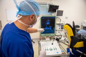
MRI fusion prostate biopsy (also known as MRI fusion TPB) is a technique that allows the urologist to accurately and comprehensively sample the prostate tissue for cell changes, which may be related to cancer or other conditions that affect the prostate gland.
Prostate biopsy is the only way to conclusively diagnose prostate cancer.
Who is suitable for an MRI fusion prostate biopsy?
MRI fusion prostate biopsy is indicated for patients that:
- Have a nodule or other abnormality on the prostate gland, that has been detected during a digital rectal examination (DRE)
- Have elevated PSA (prostate-specific antigen) levels indicated on their blood test
- Have an elevated PSA level, but an ultrasound-guided biopsy has shown negative for prostate cancer
- Have been diagnosed with prostate cancer in the past, as part of their ongoing monitoring.

What are the advantages of having an MRI fusion prostate biopsy?
MRI fusion prostate biopsy offers patients and doctors a number of advantages. It allows a large number of tissue samples to be collected accurately and efficiently from the entire prostate gland. This allows the urologist to confirm or rule out a diagnosis of significant prostate cancer. In cases where prostate cancer has previously been diagnosed, it allows the urologist to monitor for any progression.
Other conditions of the prostate are also often diagnosed. These include conditions such as BPH (benign prostatic hyperplasia), inflammation and infection of the prostate, and other non-cancerous changes.
There is no rectal wall needle insertion with MRI fusion transperineal prostate biopsy as per transrectal biopsy, and therefore the risk of infection is significantly lower.
How is an MRI fusion prostate biopsy performed?
An MRI fusion prostate biopsy is performed under general anaesthetic. Before the procedure can take place, the patient will have previously had an MRI scan of their prostate.
During the MRI fusion prostate biopsy – which typically takes 30-45 minutes – the surgeon will insert an ultrasound probe into the rectum. The live ultrasound image is ‘fused’ with the previously taken MRI scan image. Together, the surgeon is able to identify particular areas of interest on the prostate. A special biopsy grid with holes 5mm apart is positioned against the patient’s skin, behind the testicles, at the perineum. The biopsies are collected from the areas that the surgeon needs to sample. These biopsy samples are then sent for laboratory analysis under a microscope.
What can a patient expect following an MRI fusion prostate biopsy?
Patients who have an MRI fusion biopsy can expect to go home on the same day following their procedure. It is common to have some light bleeding in the perineal/biopsy area, and this can be managed very easily with gentle pressure. Typically, there may be some discomfort in the area for 1-2 days following the procedure. Some patients will notice a small amount of blood in their urine or ejaculate in the days following the procedure, but this usually resolves within a week (can rarely last longer).
Rarely, in some patients following MRI fusion prostate biopsy, a temporary inability to pass urine (urine retention) occurs. This can be fixed with the temporary placement of a urinary catheter.
MRI fusion prostate biopsy procedure outcomes:
In the vast majority of patients who undergo MRI fusion prostate biopsy, recovery time is very brief. Within just a couple of days, most patients find that they are comfortable to resume normal daily activities.
The samples collected during the MRI fusion prostate biopsy are analysed by the pathology lab, and these results are usually available for review by your urologist at your next appointment in 1-2 weeks.
MRI fusion prostate biopsy is a useful diagnostic procedure that helps to accurately diagnose prostate cancer, and to distinguish between cancer and other conditions that affect the prostate gland in men. Your urologist can then initiate the appropriate treatment plan for your individual condition, if required.
